The words sound similar, but they are different conditions.
eCollection 2023 Jan. Nour Eldine M, Alhousseini M, Nour-Eldine W, Noureldine H, Vakharia KV, Krafft PR, Noureldine MHA. Background discontinuity with interburst intervals of more than 30s is associated with a 100% probability of severe neurologic disability or death (Menache et al., 2002). Although the duration and severity of brain ischemia is often difficult to determine, clinicians are often faced with difficult issues related to predicting outcome related to awakening and long-term neurological deficits after the arrest. It may be best for children who continue to have seizures after taking two or more anti-seizure medications to get a referral to a Comprehensive Epilepsy Center to discuss other treatment options. The current therapeutic interventions are extremely limited in improving neonatal outcomes. Hypoxic ischemic injury in adults occurs mostly as a result of cerebral hypoperfusion following cardiac arrest, respiratory failure, drowning etc. Before damage to nerve cells by excessive stimulation by glutamate).
Hypothermia and systemic supportive care form the cornerstone of therapy for HIE.
The grey matter structures are affected. WebHypoxic ischemic encephalopathy (HIE) results from hypoxia or ischemia to the cerebral circulation, resulting in variable inflammation, injury, or death of neural tissues of the brain.
Gofton, in Encyclopedia of the Neurological Sciences (Second Edition), 2014. Anoxic spreading depolarization in the neonatal rat cortex. 2022 Dec 28;22(1):739. doi: 10.1186/s12887-022-03606-6. Flowchart: F21.11-3-V10-R26. WebHypoxic-ischemic encephalopathy (HIE) is a leading cause of neonatal brain injury, morbidity, and mortality globally. Journal of Neuroradiology. Which Ones? (2009) ISBN:0750675373. 2006 Feb;24(1):123-32. doi: 10.1016/j.ncl.2005.11.001. Conclusions and relevance: The similarities with periventricular leukomalacia of very premature infants are apparent.
Administration of bone marrow-derived mononuclear cells contributed to the reduction of hypoxic-ischemic brain injury in neonatal rats.
In addition, it is important to pay attention to the childs overall development and management of other associated medical problems. Unable to load your collection due to an error, Unable to load your delegates due to an error. Abnormal brain oxygen homeostasis in an animal model of liver disease. Webhypoxic-ischemic brain injury is diffuse brain damage due to reduced blood flow (ischemia) and/or reduced oxygen supply or utilization (hypoxia) to brain tissue; cardiac arrest is the
In bursts, we usually observe asynchronous and asymmetric sharp and slow waves, sometimes with multifocal or focal sharp waves. In the present study, we investigate the Diffusion-weighted MR imaging is the earliest imaging modality to become positive, usually within the first few hours after a hypoxic-ischemic event due to early cytotoxic edema. It happens when your brain doesnt get enough oxygen, which leads to brain damage. Few studies on the consequences following newborn hypoxic-ischemic encephalopathy (NHIE) assess the risk of mood disorders (MD), although these are prevalent after ischemic brain injury in adults.
Severe and prolonged interruption of placental blood flow will ultimately lead to asphyxia, the biochemical process characterized by worsening hypoxia, hypercarbia, and acidosis.
The relative risk reduction was 24%, with number needed to treat of 7 in moderate or severe HIE.
2010 Apr;10(2):60-6; quiz 67-8. doi: 10.1097/ANC.0b013e3181d54b30. Question 20 from the second paper of 2022 asked the candidates to list CT and MRI features of severe hypoxic ischaemic encephalopathy. Note: Hypoxia = blood decreased oxygen carrying capacity, [1] e.g. Infant hypoxic-ischemic encephalopathy, or HIE, is a serious birth injury caused by a lack of blood flow (ischemia) and oxygen (hypoxia) to a babys brain. Saunders. Bethesda, MD 20894, Web Policies
HIE can lead to brain damage and, in some babies, to a disability like cerebral palsy.
The reason for this predilection is not entirely clear, but it has been suggested that the relative immaturity of Purkinje cells (which are normally exquisitely sensitive to ischemic damage) in neonates somehow protects the cerebellar cortex 1. WebHypoxic ischemic encephalopathy is a neonatal brain injury that results from fetal oxygen and blood deprivation around the time of labor and delivery. MeSH
Table 14.2 gives an overview of the predictive values of EEG in the first 3 days of life for short- and long-term outcomes. . HIE can be deadly.
Smart Grocery Shopping When You Have Diabetes, Surprising Things You Didn't Know About Dogs and Cats. Most often it involves the cortex fairly diffusely along with the deep grey matter structures, particularly basal ganglia, however, many other patterns are observed 4: The predominance of grey matter injury is due to its high metabolic requirement for oxygen and glucose to supply a large number of synapses.
10.1159/000455836; Sato Y, Ueda K, Kondo T, et al. Both the reversal sign and the white cerebellum sign indicate severe injury and a poor neurologic outcome 1,3. Irene A.G. Roberts, Subarna Chakravorty, in Platelets (Third Edition), 2013, Hypoxic ischemic encephalopathy (HIE), characterized by fetal distress, fetal and neonatal acidemia, low Apgar scores at birth, and neonatal encephalopathy, is a common precipitant of neonatal thrombocytopenia. Hypoxic-ischemic cerebral injury occurs at any age, although the etiology is significantly different: Patients typically present to hospital following an acute event (near-drowning, asphyxia, cardiac/respiratory arrest).
Epilepsy centers provide you with a team of specialists to help you diagnose your epilepsy and explore treatment options.
Faan ARMD, Resnick SJ.
The topography of the neuronal injury depends in part on the severity and temporal characteristics of the insult and on the gestational age of the infant. doi: 10.33549/physiolres.934356. Dev Neurosci 2017;39:27386.
Methods HHS Vulnerability Disclosure, Help 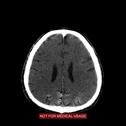
Would you like email updates of new search results? During asphyxia, not only the brain but also many other vital organs are at risk for injury. A total of 102 articles for critical review were selected based on their relevance to the incidence of HIE, pathophysiology, neuroimaging, placental pathology, biomarkers, current systemic supportive care, hypothermia, and emerging therapies for HIE and were reviewed by both of us. Careers.
myocardial infarction, drowning). 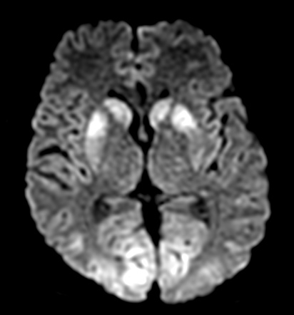 2. In this model the intranasal application of mouse MSCs improves sensorimotor function and decreases lesion size, if administered in a dose of 0.5 or 1.0 106 cells per pup.70 A smaller dose was ineffective. Unable to load your collection due to an error, Unable to load your delegates due to an error.
Strokes are one of the leading causes of mortality and chronic morbidity in the world, yet with only limited successful interventions available at present. WebAbstract: Hypoxic-ischemic encephalopathy (HIE), is one of the most frequent and dramatic urgency found in neurological brain diseases of adults. 
Finally, the term encephalopathy refers to brain dysfunction. WebIn adult population, the most common causes of hypoxic-ischemic encephalopathy are cardiac arrest or cerebrovascular disease with secondaty hypoxemia. Clinical assessment is not reliable and ancillary investigations, particularly imaging and EEG, are needed to understand the The https:// ensures that you are connecting to the
It is caused by a myriad of aetiologies, including cardiac arrest, hypotension, trauma, drowning and asphyxiation.
WebGeneral. Epub 2022 Mar 1. WebQueensland Clinical Guideline: Hypoxic ischaemic encephalopathy (HIE) Refer to online version, destroy printed copies after use Page 3 of 34 . Hypoxic-ischemic encephalopathy (HIE) resulting from asphyxia is the most common cause of neonatal brain damage and results in significant neurological sequelae, Babies with hypoxic ischemic encephalopathy (HIE) may have seizures within hours of birth. Radiographics. not neonates), also known as global hypoxic-ischemic injury, is seen in many settings and
Seizures may be very subtle in many HIE cases. Practice Essentials Perinatal asphyxia, more appropriately known as hypoxic-ischemic encephalopathy (HIE), is characterized by clinical and laboratory evidence of
Hypoxic-ischemic encephalopathy: Damage to cells in the central nervous system (the brain and spinal cord) from inadequate oxygen. However, in other cases, seizures may recur weeks to years later, and range in severity. Quick Answer.
In our series of patients with hypoxic-ischemic injury, we observed a total of seven MR imaging patterns. However, once antiseizure medications are administered, up to 58% of treated neonates exhibit electroclinical uncoupling, in which the clinical signs of their seizures vanish despite the persistence of subclinical electrographic seizures. 8600 Rockville Pike Under What Conditions? Programs Briefs | Epilepsy Foundation, Discrimination in Federally Funded Programs Briefs, First Responders and Seizure Management Briefs, Resources and Seizure Action Plans for Summer Camp, Explaining Epilepsy to Friends and Family, Epilepsy Foundation Individual and Family Services, About Research and Funding at Epilepsy Foundation, The Epilepsy Learning Healthcare System (ELHS), Access the Rare Epilepsy Network Registry, #AimForZero: Striving Toward a Future Free from Sudden Unexpected Death in Epilepsy, Advocacy: Access Prescription Medications, Advocacy: Affordable Comprehensive Health Coverage, Teens Speak Up! With the improvement in prehospital emergency systems, 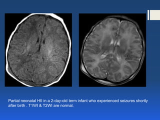 and transmitted securely. Judicious fluid management is also necessary in this population at risk for renal injury and oliguria. Already in the first hours of life, EEG background changes in HIE provide important information on the functional extent of the global brain injury and have a reliable early prognostic value. Hypoxic-ischemic brain injury most often results from insults such as cardiac arrest, vascular catastrophe, poisoning (such as carbon monoxide intoxication or drug overdose), or head trauma.
{"url":"/signup-modal-props.json?lang=us"}, Di Muzio B, Yap J, Baba Y, et al. The https:// ensures that you are connecting to the
Those changes lead to an altered mental state, leaving you confused and not acting like you usually do. Objective: A routine 1h EEG recording may miss this transition, as neonatal ultradian sleep rhythm lasts 1h (Ladanyi et al., 2004).
1.